Embark on a journey into the intricate world of cell division with our comprehensive guide to diagrama de mitosis y meiosis. From the fundamental differences between mitosis and meiosis to the fascinating stages of each process, this exploration unveils the secrets of cell reproduction with engaging prose and illuminating visuals.
Unravel the mysteries of chromosomes, centrioles, crossing over, and independent assortment as we delve into the intricacies of mitosis and meiosis, revealing their profound impact on growth, repair, and the perpetuation of life.
Mitosis and Meiosis Overview

Mitosis and meiosis are two essential cell division processes in living organisms. Mitosis produces two genetically identical daughter cells, while meiosis results in four genetically diverse gametes. Understanding the differences between these processes is crucial for comprehending cellular biology and genetics.
Mitosis occurs in somatic cells and is responsible for growth, tissue repair, and asexual reproduction. It involves four distinct phases: prophase, metaphase, anaphase, and telophase. During prophase, the chromatin condenses into visible chromosomes, and the nuclear envelope breaks down. In metaphase, the chromosomes align at the cell’s equator.
Anaphase follows, where sister chromatids separate and move to opposite poles of the cell. Finally, in telophase, two new nuclear envelopes form around the separated chromosomes, and the cell membrane pinches in the middle, resulting in two identical daughter cells.
Meiosis
Meiosis, on the other hand, is a specialized form of cell division that occurs in reproductive cells, such as eggs and sperm. Its primary function is to produce haploid gametes, which contain half the number of chromosomes as the parent cell.
Meiosis involves two successive divisions, known as meiosis I and meiosis II, each with its own unique phases.
Meiosis I begins with prophase I, during which homologous chromosomes pair up and undergo a process called crossing over, where genetic material is exchanged. The chromosomes then align at the cell’s equator in metaphase I, followed by separation and movement to opposite poles in anaphase I.
Diagrams of mitosis and meiosis are essential for understanding cell division. These diagrams can help us visualize the complex processes involved in cell reproduction. As Isaac Asimov wrote in his short story “The Last Answer” , “The most exciting phrase to hear in science, the one that heralds new discoveries, is not ‘Eureka!’ (I found it!) but ‘That’s funny…'”.
Similarly, diagrams of mitosis and meiosis can help us identify anomalies and ask questions that lead to new insights into cell biology.
In telophase I, two haploid daughter cells are formed, each containing one set of unreplicated chromosomes.
Meiosis II follows a similar pattern to mitosis, with prophase II, metaphase II, anaphase II, and telophase II. However, during meiosis II, the chromosomes do not replicate again, resulting in four haploid gametes with unique genetic combinations.
Mitosis in Detail

Mitosis is a type of cell division that results in two daughter cells that are genetically identical to the parent cell. It is a continuous process, but for the sake of explanation, it can be divided into four distinct stages: prophase, metaphase, anaphase, and telophase.During
prophase, the chromosomes become visible and the nuclear envelope begins to break down. The centrioles, which are responsible for organizing the spindle fibers, begin to move to opposite poles of the cell. In metaphase, the chromosomes line up in the center of the cell.
The spindle fibers attach to the chromosomes and begin to pull them apart. In anaphase, the chromosomes continue to be pulled apart until they reach opposite poles of the cell. In telophase, the spindle fibers disappear and the nuclear envelope reforms around each of the two daughter cells.
Role of Chromosomes and Centrioles in Mitosis
Chromosomes are structures that contain the cell’s genetic material. During mitosis, the chromosomes are duplicated so that each daughter cell receives a complete set of chromosomes. Centrioles are structures that are involved in organizing the spindle fibers. The spindle fibers are responsible for pulling the chromosomes apart during mitosis.
Meiosis in Detail: Diagrama De Mitosis Y Meiosis

Meiosis is a specialized cell division that produces haploid gametes, such as sperm and eggs. It involves two rounds of division, known as meiosis I and meiosis II, resulting in four haploid daughter cells from a single diploid parent cell.
Stages of Meiosis
Meiosis I
- Prophase I:Chromosomes condense and homologous chromosomes pair up, exchanging genetic material through a process called crossing over.
- Metaphase I:Homologous chromosome pairs line up at the equator of the cell.
- Anaphase I:Homologous chromosomes separate and move to opposite poles of the cell.
- Telophase I:Two haploid daughter cells are formed, each containing one set of chromosomes.
Meiosis II
- Prophase II:Chromosomes condense again.
- Metaphase II:Chromosomes line up at the equator of each daughter cell.
- Anaphase II:Sister chromatids separate and move to opposite poles of each cell.
- Telophase II:Four haploid daughter cells are formed, each containing one set of chromosomes.
Crossing Over and Independent Assortment
Crossing over is a key event in meiosis that increases genetic diversity. During crossing over, homologous chromosomes exchange genetic material, resulting in new combinations of alleles. Independent assortment is another mechanism that contributes to genetic diversity. It occurs during meiosis I when homologous chromosomes line up randomly at the equator, ensuring that each daughter cell receives a unique combination of maternal and paternal chromosomes.
Comparison of Mitosis and Meiosis

Mitosis and meiosis are two distinct types of cell division that serve different purposes in the life cycle of eukaryotic organisms. Mitosis is responsible for growth and tissue repair, while meiosis is involved in sexual reproduction.
The key differences between mitosis and meiosis are summarized in the following table:
Table of Key Features
| Feature | Mitosis | Meiosis |
|---|---|---|
| Chromosome number | Diploid (2n) | Haploid (n) |
| Daughter cells produced | Two identical daughter cells | Four haploid daughter cells |
| Purpose | Growth and tissue repair | Sexual reproduction |
Applications of Mitosis and Meiosis

Mitosis and meiosis are essential processes in the life cycle of organisms. Mitosis is responsible for growth, repair, and asexual reproduction, while meiosis plays a crucial role in sexual reproduction and genetic diversity.
Applications of Mitosis
Mitosis is a type of cell division that produces two genetically identical daughter cells from a single parent cell. It is essential for the following:
- Growth:Mitosis allows organisms to increase in size by producing new cells.
- Repair:When cells are damaged or die, mitosis replaces them with new, functional cells.
- Asexual reproduction:In some organisms, mitosis is the only means of reproduction. Each daughter cell produced by mitosis can develop into a new individual that is genetically identical to the parent.
Applications of Meiosis
Meiosis is a type of cell division that produces four genetically unique daughter cells from a single parent cell. It is essential for the following:
- Sexual reproduction:Meiosis produces gametes (eggs and sperm) that fuse during fertilization to create a genetically diverse offspring.
- Genetic diversity:The process of meiosis involves genetic recombination, which shuffles the genetic material of the parent cells. This results in the production of offspring that have unique combinations of genes, contributing to genetic diversity within populations.
Illustrations and Diagrams
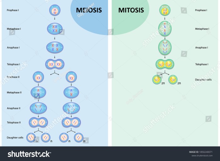
Visual representations of mitosis and meiosis provide a detailed understanding of the key events occurring during these cellular processes.
The following illustrations present the stages of mitosis and meiosis, accompanied by descriptive captions explaining the critical events.
Mitosis, Diagrama de mitosis y meiosis
- Prophase:Chromosomes become visible, and the nuclear envelope breaks down.
- Metaphase:Chromosomes align at the equator of the cell.
- Anaphase:Sister chromatids separate and move to opposite poles of the cell.
- Telophase:Two new nuclear envelopes form around the separated chromosomes, and the cell divides into two daughter cells.
Meiosis
- Prophase I:Homologous chromosomes pair up and exchange genetic material (crossing over). Tetrads form.
- Metaphase I:Homologous chromosome pairs align at the equator of the cell.
- Anaphase I:Homologous chromosomes separate and move to opposite poles of the cell.
- Telophase I:Two daughter cells are formed, each containing one chromosome from each homologous pair.
- Prophase II:Chromosomes become visible again, and the nuclear envelope breaks down.
- Metaphase II:Chromosomes align at the equator of the cell.
- Anaphase II:Sister chromatids separate and move to opposite poles of the cell.
- Telophase II:Four new nuclear envelopes form around the separated chromosomes, and the cell divides into four daughter cells.
Key Questions Answered
What is the primary distinction between mitosis and meiosis?
Mitosis produces two identical daughter cells with the same number of chromosomes as the parent cell, while meiosis produces four genetically diverse daughter cells with half the number of chromosomes as the parent cell.
How many phases are there in mitosis?
Mitosis consists of four phases: prophase, metaphase, anaphase, and telophase.
What is the role of crossing over in meiosis?
Crossing over is a genetic recombination process that occurs during meiosis, where homologous chromosomes exchange genetic material, resulting in increased genetic diversity.