Embark on a journey into the intricate world of human brain cranial nerves coloring, a technique that illuminates the anatomy and function of the nervous system. Delve into the depths of the human brain, unraveling the complexities of its structure and the vital roles played by the cranial nerves.
This comprehensive guide explores the intricacies of brain anatomy, the identification and pathways of cranial nerves, and the principles behind various staining techniques. Discover how these techniques enhance our understanding of brain development, aid in the diagnosis of neurological disorders, and guide surgical interventions.
Human Brain Anatomy
The human brain, housed within the protective cranial cavity, is a complex and intricate organ responsible for controlling various bodily functions, including cognition, emotions, and movement. Understanding its anatomy is crucial for comprehending its role in human physiology.
The brain can be broadly divided into three major regions: the forebrain, midbrain, and hindbrain. The forebrain comprises the cerebral hemispheres, which are responsible for higher-level functions such as reasoning, memory, and language. The midbrain connects the forebrain to the hindbrain and is involved in motor control and sensory processing.
The hindbrain includes the cerebellum, which coordinates movement and balance, and the medulla oblongata, which controls vital functions such as breathing and heart rate.
The brain is protected by three layers of membranes known as the meninges. The outermost layer, the dura mater, is a tough, fibrous membrane. The middle layer, the arachnoid mater, is a delicate web-like membrane. The innermost layer, the pia mater, is a thin membrane that closely adheres to the surface of the brain.
Between the arachnoid mater and pia mater lies the subarachnoid space, which contains cerebrospinal fluid that cushions and nourishes the brain.
Cranial Nerves

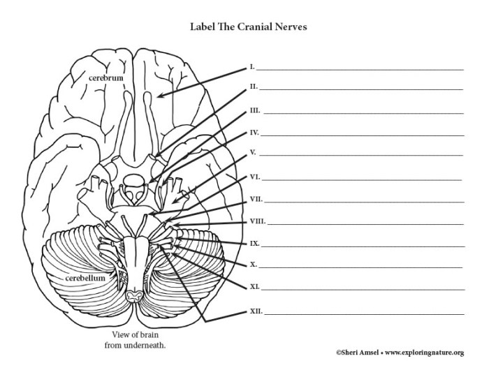
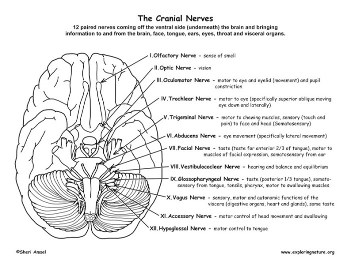
The cranial nerves are twelve pairs of nerves that emerge directly from the brain and innervate various structures in the head and neck. They play a crucial role in sensory and motor functions, including vision, hearing, taste, smell, and muscle movement.
Each cranial nerve has a specific name and number, as well as a unique pathway and distribution. The olfactory nerve (I) is responsible for the sense of smell, while the optic nerve (II) transmits visual information from the eyes to the brain.
The oculomotor nerve (III), trochlear nerve (IV), and abducens nerve (VI) control eye movements. The trigeminal nerve (V) provides sensory innervation to the face, while the facial nerve (VII) controls facial muscles. The vestibulocochlear nerve (VIII) is responsible for hearing and balance.
The glossopharyngeal nerve (IX) and vagus nerve (X) innervate structures in the throat and neck, including the tongue and larynx. The accessory nerve (XI) innervates the sternocleidomastoid and trapezius muscles, while the hypoglossal nerve (XII) controls tongue movement.
Cranial nerve examinations are essential in diagnosing neurological disorders. By assessing the function of each cranial nerve, clinicians can identify damage or dysfunction in specific regions of the brain or along the nerve pathways.
Coloring Techniques for the Brain and Cranial Nerves
Coloring techniques are essential tools in neuroanatomy, allowing researchers and clinicians to visualize and study the brain and cranial nerves in detail. These techniques involve staining specific structures or components within the nervous tissue to enhance their visibility and contrast under a microscope.
One commonly used staining technique is Nissl staining, which utilizes basic dyes to highlight the Nissl bodies, which are protein-rich structures within neurons. Golgi staining, on the other hand, uses silver salts to selectively stain a small subset of neurons, providing a detailed view of their morphology and connections.
Immunohistochemistry is another powerful technique that utilizes antibodies to visualize specific proteins within the brain tissue, allowing researchers to study the distribution and expression of various proteins.
To prepare brain tissue for staining, it is typically fixed in a preservative solution, such as formalin, to prevent decomposition. The tissue is then embedded in paraffin or frozen and sectioned into thin slices using a microtome. The sections are then stained using the desired technique and mounted on glass slides for microscopic examination.
Applications of Brain and Cranial Nerve Coloring: Human Brain Cranial Nerves Coloring

Coloring techniques have a wide range of applications in studying the brain and cranial nerves. They are essential for understanding brain development, as they allow researchers to visualize the formation and organization of neural structures during embryonic and postnatal development.
Coloring techniques also play a crucial role in identifying pathological changes in the brain. By staining brain tissue from individuals with neurological disorders, researchers can visualize the presence of tumors, neurodegenerative lesions, and other abnormalities that may not be apparent through other imaging techniques.
Furthermore, coloring techniques are used in surgical procedures to guide surgeons during complex brain operations. By staining specific structures or pathways, surgeons can more accurately identify anatomical landmarks and minimize damage to surrounding tissues.
Helpful Answers
What is the purpose of staining techniques in human brain cranial nerves coloring?
Staining techniques enhance the visibility and differentiation of brain structures and cranial nerves in anatomical specimens, facilitating detailed observation and analysis.
How do staining techniques aid in the diagnosis of neurological disorders?
By highlighting pathological changes in the brain, such as tumors or neurodegenerative lesions, staining techniques assist in the accurate diagnosis and characterization of neurological disorders.
What are the applications of human brain cranial nerves coloring in surgical procedures?
During complex brain surgeries, coloring techniques provide visual guidance to surgeons, enabling precise navigation and minimizing damage to delicate brain structures.